Computed Tomography (CT, X-Ray CT, CAT)

What
Computed tomography (CT) is a medical imaging technique that produces cross-sectional images and 3D volume renderings of the body's internal structures using X-rays. This technique is also called X-ray computed tomography (X-ray CT) and computerized axial tomography (CAT). The word tomography refers to cutting or slicing.
Why
Computed tomography is often used as an imaging tool for the following purposes:
Looking at the head to detect tissue death due to local lack of oxygen, tumors, calcifications, bleeding, and bone trauma.
Looking at the lungs to detect defects.
Looking at the pulmonary arteries to diagnose pulmonary embolism. This technique is known as a CT pulmonary angiogram.
Looking at the coronary arteries to diagnose coronary artery disease. This technique is known as a CT coronary angiogram.
Looking at the abdomen to determine the stage of cancer, diagnose abdominal diseases, and diagnose abdominal pain.
Looking at complex bone fractures, ligament injuries, and dislocations.
Screening for diseases such as colon cancer or heart disease in higher-risk individuals.
How
Computed tomography functions using the same fundamental technology as projectional radiography. The main difference is that in computed tomography the X-ray source and image detector are spinning axially around the patient as the patient moves longitudinally through the X-rays.
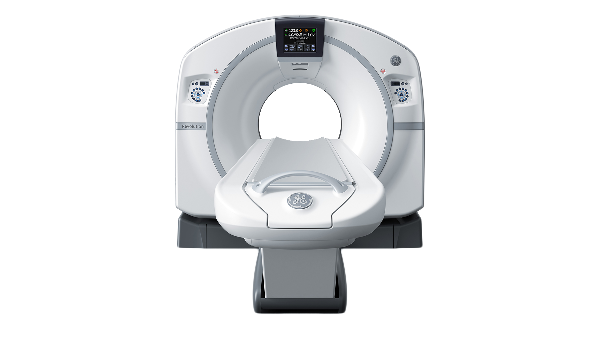
A typical computed tomography system consists of the following main components:
Rotating Gantry: This is a circular structure to which the X-ray tube and image detector are mounted. The gantry rotates continuously in order to image the patient from many different angles.
X-Ray Tube: This is what produces the X-rays. This is mounted to the rotating gantry opposite of the image detector.
Patient: The X-rays go through the patient and some of the radiation is absorbed. Denser structures such as bone absorb more X-rays than softer structures.
Table: The table moves longitudinally through the rotating gantry.
Image Detector: This is what collects the X-rays that went through the patient. This is mounted to the rotating gantry opposite of the X-ray tube.
User Interface: This is how the user operates the system.
Processor: This allows the user to produce a stack of cross-sectional images and produce 3D renderings.
Radiocontrast Agent: Computed tomography is often performed using a radiocontrast agent to improve visibility of the target structures. Common radiocontrast agents include iodine, barium, air, and carbon dioxide.

Components of a Typical Computed Tomography System
Who
Some of the main companies who make computed tomography systems include:
GE (Revolution, Optima)
Philips (IQon, Incisive, Ingenuity, Big Bore)
Siemens (SOMATOM)
Canon, Toshiba (Aquilon)
Hitachi (SCENARIA, Supria)
References
http://www3.gehealthcare.com/en/products/categories/computed_tomography
http://www.usa.philips.com/healthcare/solutions/computed-tomography/ct-scanners
http://www.hitachimed.com/products/ComputedTomography/?WT.ac=med_mg_pro_ct
https://en.wikipedia.org/wiki/Computed_tomography_of_the_heart