Magnetic Resonance Imaging (MRI, NMRI, MRT)
Video

What
Magnetic resonance imaging (MRI) is a medical imaging technique that uses magnetic fields, radio waves, and field gradients to produce images of the body's internal structures. This imaging technique is also called nuclear magnetic resonance imaging (NMRI) and magnetic resonance tomography (MRT). Unlike computed tomography, magnetic resonance imaging does not expose the patient to ionizing radiation.
Why
Magnetic resonance imaging is often used for the following purposes:
Looking at the brain and brain stem to investigate neurological cancers, abnormalities, and disorders.
Looking at the heart and other parts of the cardiovascular system. This practice is known as cardiovascular MRI (CMR). Measuring flow velocities of blood throughout the body can be done with phase contrast MRI (PC-MRI).
Looking at arteries to identify stenosis or aneurysms. This practice is known as magnetic resonance angiography (MRA).
Looking at structures within the musculoskeletal system such as the spine, joints, and soft tissue tumors.
Looking at the liver, pancreas, bile ducts, and the gastrointestinal tract.
Staging and follow-up imaging of rectal and prostate cancer and diagnosing, staging, and follow-up imaging of other tumors.
Guiding instruments in three dimensions during minimally-invasive interventional procedures. This practice is known as interventional MRI (IMRI).
Guiding the application of high-intensity focused ultrasound (HIFU) to ablate diseased tissue. This practice is known as magnetic resonance guided focused ultrasound (MRgFUS).
Imaging while performing surgery to guide and confirm the success of the procedure. This practice is known as intraoperative MRI.
Measuring brain activity. This practice is known as functional MRI (fMRI).
Measuring the diffusion of water molecules to diagnose conditions such as stroke and neurological disorders such as multiple sclerosis. This practice is known as diffusion-weighted MRI (DW-MRI).
Measuring levels of metabolites to diagnose metabolic disorders, particularly those in the brain. This practice is known as magnetic resonance spectroscopy (MRS).
Visualizing the cellular function of molecular processes using biomarkers for the diagnosis of cancer as well as neurological and cardiovascular diseases. This practice is a form of molecular imaging.
How
Magnetic resonance imaging relies on nuclear magnetic resonance, a phenomenon in which nuclei in a magnetic field absorb and re-emit electromagnetic radiation.
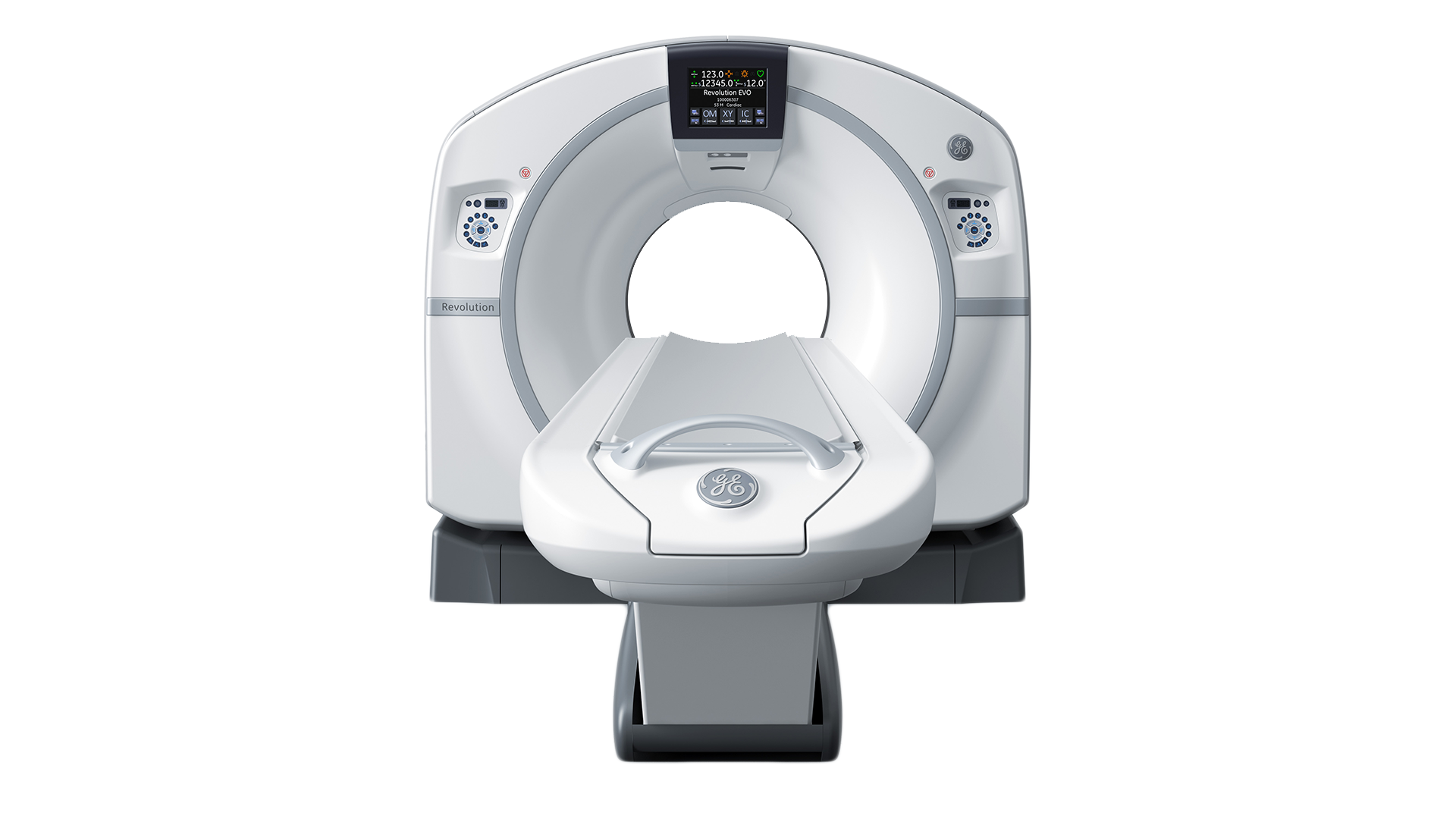
A typical magnetic resonance imaging system consists of the following main components:
Magnet: This creates a powerful and uniform magnetic field which points straight through the bore.
Patient: The patient's body consists largely of water. The magnetic field causes the hydrogen protons in the water to polarize and point toward either the patient's head or feet. Pairs of protons pointing in opposite directions cancel each other out while a much smaller number of unmatched protons do not.
Radiofrequency Coil: This applies an intense pulse of radiofrequency energy which causes the unmatched protons to spin at a specific frequency and in a certain direction. Once the pulse is turned off, the hydrogen protons slowly re-align within the magnetic field. As they do, the protons re-emit the energy that they absorbed from the pulse and this signal is detected by the same radiofrequency coil.
Gradient Coils: These magnets are switched on and off rapidly in a controlled sequence to locally perturb the main magnetic field. This enables spatial encoding of the signal such that thin slices can be imaged in any orientation and location within the body.
Processor: This utilizes a Fourier transform to convert the complex time-domain signal into a frequency-domain signal and ultimately produce 2D and 3D images.
Table: This is what the patient lays on.
Contrast Agent: Magnetic resonance imaging can be performed with contrast agents which affect the relaxation times of atoms. The most common contrast agents are gadolinium-based.
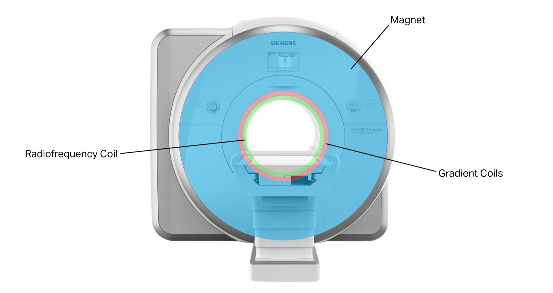
Components of a Typical Magnetic Resonance Imaging System
Who
Some of the main companies who make magnetic resonance imaging systems include:
GE (SIGNA)
Siemens (MAGNETOM)
Philips (Ingenia)
Canon, Toshiba (Vantage)
Hitachi (Oasis, Echelon, Legacy)
References
http://www3.gehealthcare.com/en/products/categories/magnetic_resonance_imaging
https://usa.healthcare.siemens.com/magnetic-resonance-imaging
http://www.usa.philips.com/healthcare/solutions/magnetic-resonance
http://www.hitachimed.com/products/mri/?WT.ac=med_mg_pro_mri
https://en.wikipedia.org/wiki/Magnetic_resonance_imaging_of_the_brain
https://en.wikipedia.org/wiki/Cardiac_magnetic_resonance_imaging
https://en.wikipedia.org/wiki/Functional_magnetic_resonance_imaging
https://en.wikipedia.org/wiki/Phase_Contrast_Magnetic_Resonance_Imaging
https://en.wikipedia.org/wiki/Physics_of_magnetic_resonance_imaging
https://en.wikipedia.org/wiki/Interventional_magnetic_resonance_imaging
https://en.wikipedia.org/wiki/Magnetic_resonance_angiography
https://en.wikipedia.org/wiki/In_vivo_magnetic_resonance_spectroscopy
https://en.wikipedia.org/wiki/High-intensity_focused_ultrasound