Gastroenterology Endoscopes
Video

What
Gastroenterology endoscopes are flexible or rigid tubes inserted into the gastrointestinal tract via the mouth or anus to perform a variety of diagnostic and therapeutic procedures. At a minimum, endoscopes enable the user to see within the gastrointestinal tract; however, most endoscopes also enable other endoscopic devices to be inserted through a working channel to perform physical procedures.
Why
Endoscopes are typically used for the following diagnostic and therapeutic purposes within and adjacent to the gastrointestinal tract:
Visually examining and diagnosing abnormalities and diseases such as polyps, ulcers, bleeding, inflammation, lesions, and cancer.
Imaging organs within and adjacent to the gastrointestinal tract using ultrasound. This technique is known as endoscopic ultrasound (EUS) and the endoscopes are often known as echoendoscopes. Endoscopic ultrasound is used to guide many endoscopic therapeutic procedures.
Performing biopsies using instruments inserted through the endoscope's working channel. Fine needle aspiration (FNA) is a type of biopsy used to collect samples from the gastrointestinal wall, liver, pancreas, lymph nodes, and bile ducts.
Performing therapeutic procedures using instruments inserted through the endoscope's working channel. Such procedures include polypectomy, draining pancreatic pseudocysts, dilating or stenting structures which have become too narrow, tightening structures which have become too loose, performing injection therapy, performing laser and radiofrequency ablation, and stopping bleeding.
Performing physical manipulation using instruments inserted through the endoscope's working channel. Such procedures include removing swallowed objects and placing feeding tubes.
Types
Endoscopes that are typically inserted through the mouth for use in the upper gastrointestinal tract include:
Gastroscope: This type of flexible endoscope can reach through the stomach up to the duodenum of the small bowel. Gastroscopes are used for diagnostic and therapeutic esophagogastroduodenoscopy (EGD).
Enteroscope: This type of flexible endoscope can reach through the duodenum, jejunum, and ilium of the small bowel. Enteroscopes are used for diagnostic and therapeutic enteroscopy. The small bowel is about 20 ft long and has many twists and turns. Navigation is achieved using methods such as single-balloon enteroscopy (SBE), double-balloon enteroscopy (DBE), or spiral enteroscopy.
Duodenoscope: This type of flexible endoscope is typically used to perform diagnostic and therapeutic endoscopic retrograde cholangiopancreatography (ERCP) to diagnose and treat conditions of the bile ducts and main pancreatic duct.
Choledochoscope: This type of flexible endoscope is typically used for visualization of the biliary tree when performing endoscopic retrograde cholangiopancreatography (ERCP) to diagnose and treat conditions of the bile ducts and main pancreatic duct.
Endoscopes which are typically inserted through the anus for use in the lower gastrointestinal tract include:
Colonoscope: This type of flexible endoscope can reach the distal portion of the small bowel. Colonoscopes are used for diagnostic and therapeutic colonoscopy.
Sigmoidscope: This type of flexible or rigid endoscope can reach the sigmoid colon. Sigmoidscopes are used for diagnostic and therapeutic sigmoidoscopy.
How
The distal end of the endoscope is inserted into the gastrointestinal tract and the user operates the controls on the proximal end.
Typical components at the distal end include:
Bending Section: This section can be articulated vertically and horizontally to navigate through the gastrointestinal tract.
Illumination Lens: Light produced by an external light source is transmitted via fiber optics and exits this lens. This light is then reflected and returned through the objective lens to form the image.
Objective Lens: Reflected light enters the objective lens. Fiber endoscopes transmit this light through the endoscope via fiber optics whereas video endoscopes capture the light using an imaging sensor within the distal tip.
Working Channel: This lumen allows endoscopic instruments to be inserted through the endoscope to perform procedures such as fine needle aspiration (FNA), drainage, injection, laser ablation, radiofrequency ablation, biopsy, polypectomy, and electrosurgical coagulation and cutting.
Gas, Fluid, and Suction Nozzle: This lumen allows insufflation, irrigation, and suction to optimize access and visibility within the gastrointestinal tract.
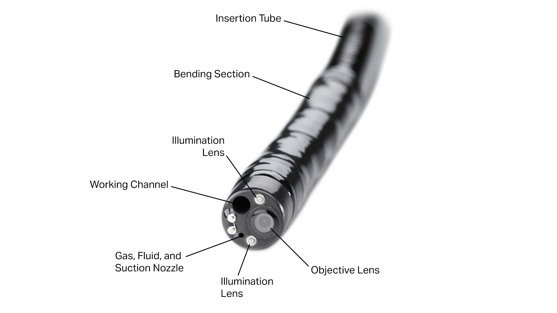
Distal Components of a Typical Flexible Gastroenterology Endoscope
Typical components at the distal end of ultrasound endoscopes include:
Ultrasound Transducer: This transmits and receives ultrasound waves using the effect of piezoelectricity. Inside the transducer, there are one or more piezoelectric crystals. When electrical pulses are applied to the crystals, they oscillate and produce ultrasound waves. When ultrasound waves are reflected back, the piezoelectric crystals oscillate and produce electrical pulses. Refer to the post on ultrasound for more information. The transducer shape determines the field-of-view. A curved linear array transducer images in a plane parallel with the axis of the endoscope. A radial array transducer images in a plane perpendicular to the axis of the endoscope.
Working Channel with Elevator: Curved linear array ultrasound endoscopes typically have a working channel with an elevator. The elevator sets the angle at which instruments exit the working channel.

Additional Distal Components of Typical Gastroenterology Ultrasound Endoscopes
Typical components at the proximal end include:
Insertion Tube: This is the long flexible section of the endoscope which is not steerable.
Vertical Deflection Control and Lock: These controls allow the bending section to be steered up and down and locked in place.
Horizontal Deflection Control and Lock: These controls allow the bending section to be steered left and right and locked in place.
Working Channel: This lumen allows endoscopic instruments to be inserted through the endoscope to perform procedures such as fine needle aspiration (FNA), drainage, injection, laser ablation, radiofrequency ablation, biopsy, and polypectomy.
Elevator Control: This control allows the angle of the elevator to be adjusted to set the angle at which instruments exit the working channel. This control is typically only found on certain types of ultrasound endoscopes.
Gas, Fluid, and Suction Controls: These controls allow for insufflation, irrigation, and suction to optimize access and visibility within the gastrointestinal tract.
Video Controls: These controls allow the image to be adjusted.
External Cord: This cord connects the endoscope to external light, gas, fluid, and suction sources and to an external display.

Proximal Components of a Typical Flexible Gastroenterology Endoscope
Who
Some of the main companies who make gastroenterology endoscopes include:
Olympus (EVIS EXERA, GF)
PENTAX (EC, 90i, 90K, ES, i10, DEC, 70K, EG, J10)
Fujifilm (ELUXEO, EC, EG, ED, EI, EN)
KARL STORZ (SILVER SCOPE)
References
http://medical.olympusamerica.com/specialty/gastroenterology
http://www.fujifilmusa.com/products/medical/endoscopy/endoscopes/index.html
https://en.wikipedia.org/wiki/Endoscopic_retrograde_cholangiopancreatography
http://www.slideshare.net/tho4497/endoscopy?next_slideshow=1