Fluoroscopy (X-Ray)
Video

What
Fluoroscopy is a medical imaging technique that produces a real-time view of the body's internal structures using X-rays. Whereas projectional radiography produces a static X-ray image, fluoroscopy produces a live X-ray video. Fluoroscopy is one of the main imaging modalities used in interventional radiology. When used for interventional procedures, fluoroscopy systems are often called interventional X-ray systems.
Why
Fluoroscopy is often used as an imaging tool for the following purposes:
To guide catheters through blood vessels, bile ducts, and urinary system.
To place devices such as stents in the body.
To look at blood vessels and organs such as the heart, a practice called angiography.
To position orthopedic implants such as artificial knees and hips.
To look at the gastrointestinal tract.
How
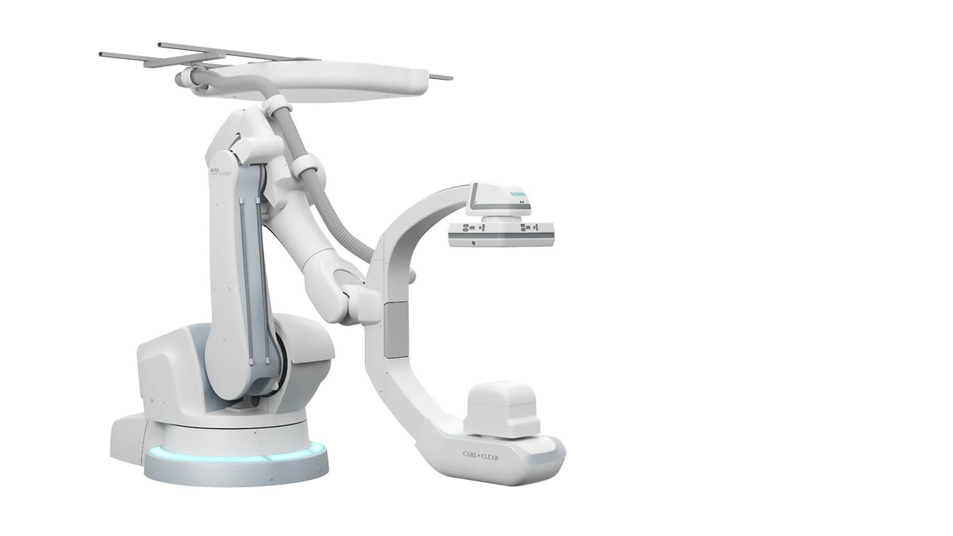
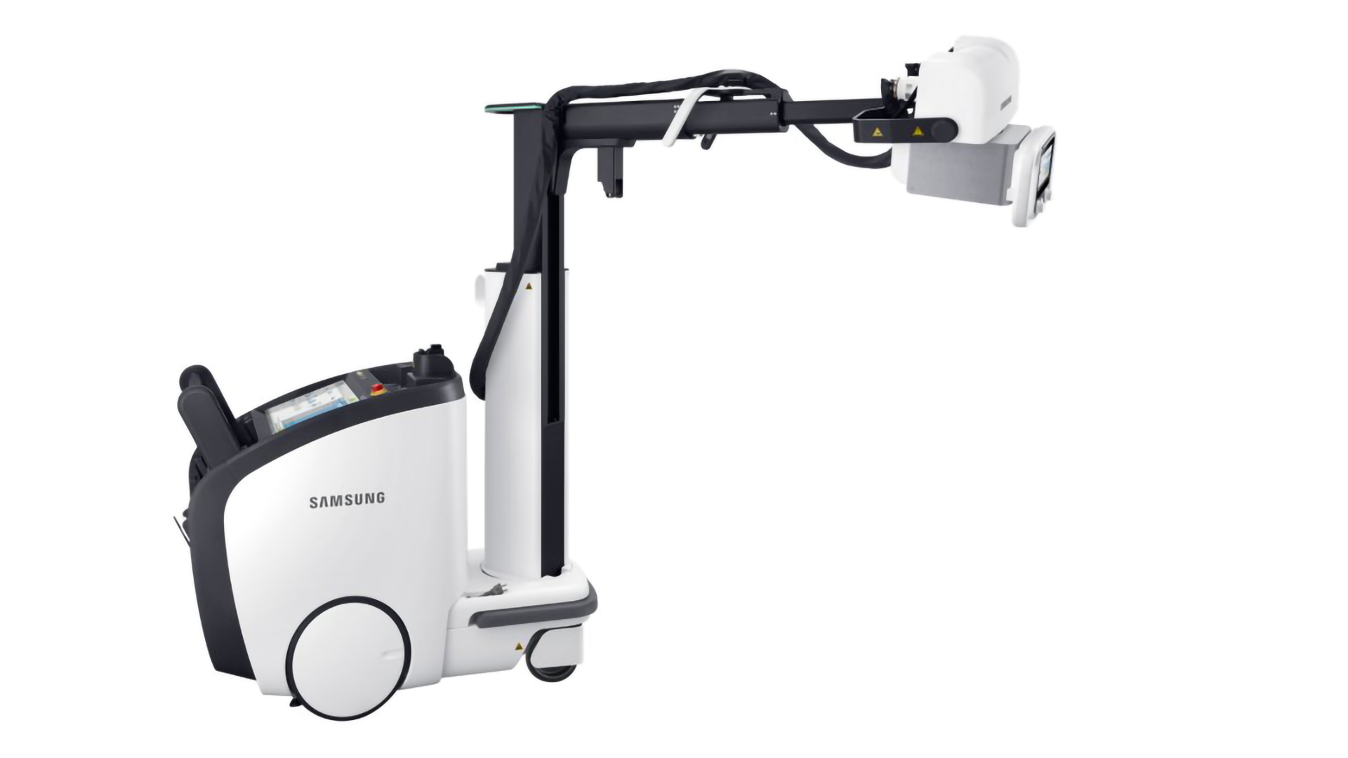
Fluoroscopy functions using the same fundamental technology as projectional radiography. A typical fluoroscopy system consists of the following main components:
X-Ray Tube: This is what produces the X-rays. An X-ray tube is a vacuum tube which applies a high voltage to accelerate electrons released from a hot cathode to an extremely high velocity. These high-energy electrons collide with a metal anode, producing X-rays.
Collimator: This focusses the X-ray beam by allowing only X-rays traveling in a certain direction to pass through.
Patient: The X-rays go through the patient and some of the radiation is absorbed. Denser structures such as bone absorb more X-rays than softer structures.
Table: This is what the patient lays on.
Bucky-Potter Grid: This may be placed between the patient and the image detector to reduce the quantity of scattered X-rays reaching the detector. This improves the contrast resolution of the image but also increases the radiation exposure for the patient.
Image Detector: This is what collects the X-rays that went through the patient to form the moving image.
User Interface: This is how the user operates the fluoroscopy system.
Display: This displays the live X-ray view.
Radiocontrast Agent: Fluoroscopy is often performed using a radiocontrast agent to improve visibility of the target structures. Common radiocontrast agents include iodine, barium, air, and carbon dioxide.

Components of a Typical Fluoroscopy System

Components of a Typical Interventional X-Ray C-Arm System

X-Ray Tubes
Who
Some of the main companies who make fluoroscopy systems include:
GE (Precision, Discovery, Innova, OEC, Uroview)
Philips (CombiDiagnost, ProxiDiagnost, Azurion, Allura, Zenition, BV Pulsera, Veradius Unity, BV Endura)
Siemens (Luminos, Artis, Cios)
Shimadzu (SONIALVISION, FLEXAVISION, FLUOROspeed, Trinias, OPESCOPE)
Canon, Toshiba (Alphenix)
Carestream (DRX)
Hitachi (DR)
References
http://www.usa.philips.com/healthcare/solutions/fluoroscopy/fluoroscopy, http://www.usa.philips.com/healthcare/solutions/interventional-xray
http://www3.gehealthcare.com/en/products/categories/radiography_and_fluoroscopy
http://shimadzu.com/med/products/fluoro/index.html, http://www.shimadzu.com/med/products/c-arm/index.html
http://www.toshibamedicalsystems.com/products/xray/rf/index.html, http://www.toshibamedicalsystems.com/products/xray/mc/index.html
http://www.hitachi.com/businesses/healthcare/products-support/xtv/index.html