Ultrasound
Video

What
Ultrasound is a medical imaging technique that uses high-frequency sound waves to produce real-time 2D and 3D views inside the body.
Why
Ultrasound is often used for the following purposes:
For monitoring development of the fetus during pregnancy. This practice is known as obstetrical sonography.
For guiding needles and cannulas into difficult vasculature.
For assessing blood flow and for diagnosing vascular diseases.
For diagnosing abnormal structure and function of the heart. This practice is known as echocardiography.
For emergency assessment of trauma.
For imaging the solid organs of the abdomen.
For imaging organs within and adjacent to the gastrointestinal tract. This technique is known as endoscopic ultrasound (EUS) and the endoscopes are often known as echoendoscopes. Endoscopic ultrasound is used to guide many endoscopic therapeutic procedures. Refer to this post on gastroenterology endoscopes for more information on endoscopic ultrasound.
For imaging structures of the neck, such as the thyroid and parathyroid glands, lymph nodes, and salivary glands.
For imaging the eyes. This practice is known as ocular ultrasonography.
For visualizing endobronchial lesions and lymph nodes prior to transbronchial needle aspiration. This practice is known as endobronchial ultrasound (EBUS).
For imaging organs of the pelvis, such as the bladder, uterus, ovaries, testicles, and prostate.
For imaging tendons, muscles, nerves, ligaments, soft tissue masses, and bone.
How
Ultrasound systems transmit high-frequency sound waves into the body and then detect the echoes that are reflected back. This technology is similar to echolocation used by bats as well as sonar used in submarines.
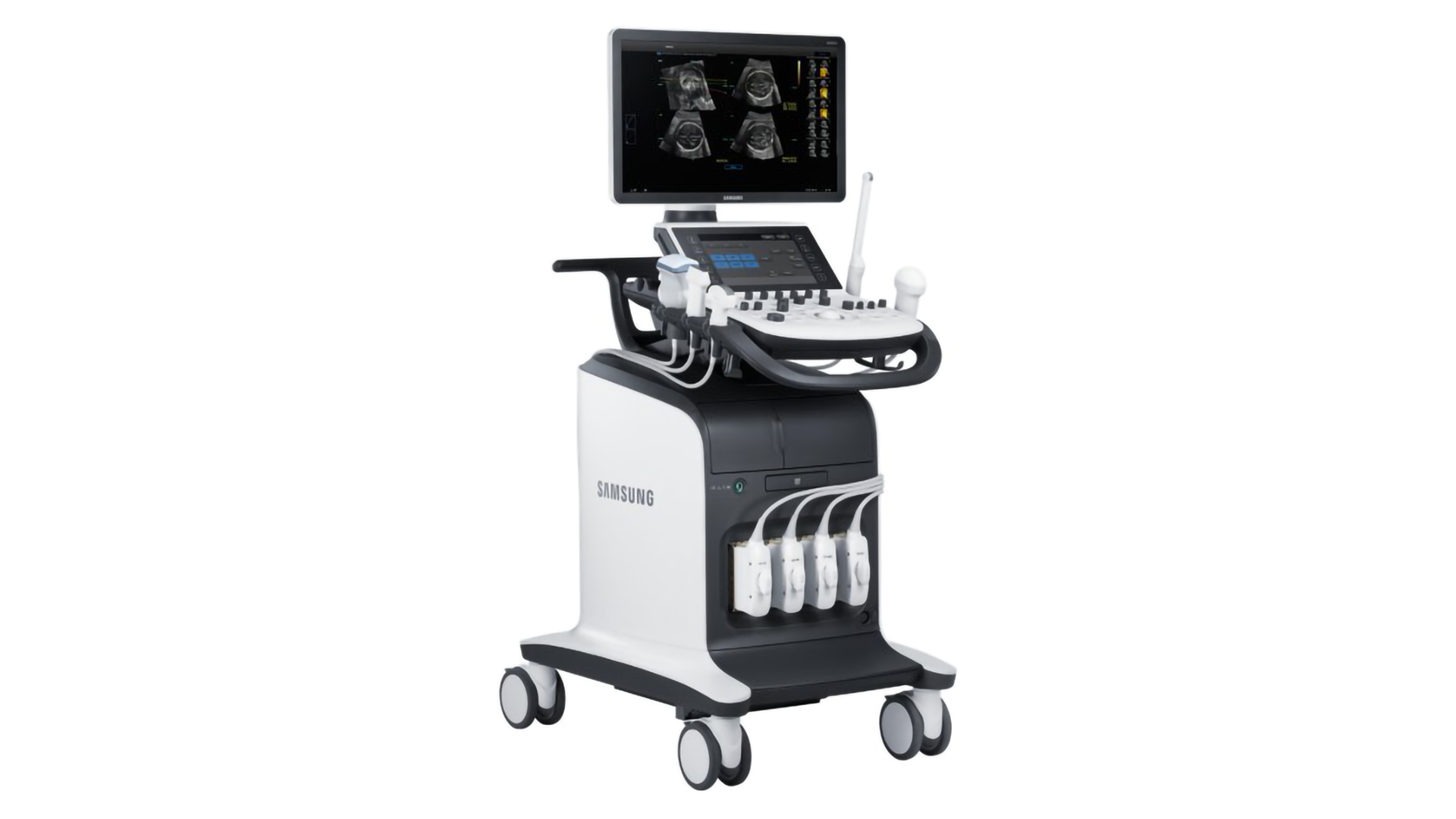
Ultrasound imaging typically involves the following main components:
Transducer Probe: The transducer probe both transmits and receives the ultrasound waves using the effect of piezoelectricity. Inside the transducer probe, there are one or more piezoelectric crystals. When electrical pulses are applied to the crystals, they oscillate and produce ultrasound waves. There is a sound-absorbing backing layer behind the crystals to eliminate back reflections and an acoustic lens in front of the crystals to focus the emitted waves. When ultrasound waves are reflected back, the piezoelectric crystals oscillate and produce electrical pulses. The face of the transducer probe is typically a rubbery material to efficiently transmit the ultrasound waves. The shape of the transducer probe determines the field-of-view.
Patient: Ultrasound waves are transmitted into the patient and come into focus at the desired depth. Some of these waves are partially reflected back to the transducer probe due to acoustic impedance changes at tissue interfaces. Each reflected wave has a measurable time delay and intensity based on the depth and composition of the tissue interface.
Processor: The processor drives the piezoelectric crystals in the transducer probe to produce the ultrasound waves. In multi-crystal probes, beamforming can be used to focus the ultrasound waves to the location of interest. Beamforming works by controlling the timing and amplitude of the waves emitted from each crystal to produce a pattern of constructive and destructive interference. The processor also receives the electrical signals back from the crystals in the transducer probe. These signals are processed to form a 2D image based on the time delay and intensity of each reflected wave. The processor can produce 3D volume renderings by combining multiple 2D scans. The processor can also measure the change in frequency of reflected ultrasound waves to calculate speed. This technique is known as Doppler ultrasound.
User Interface: This allows the operator to adjust the frequency, amplitude, and pulse duration of the ultrasound waves. Ultrasound waves are typically produced in the range of 1 to 18 MHz. Lower-frequency waves travel deeper into the body but produce lower-resolution images. Higher-frequency waves do not travel as deep but can produce higher-resolution images of smaller structures.
Display: This displays the 2D or 3D ultrasound image.
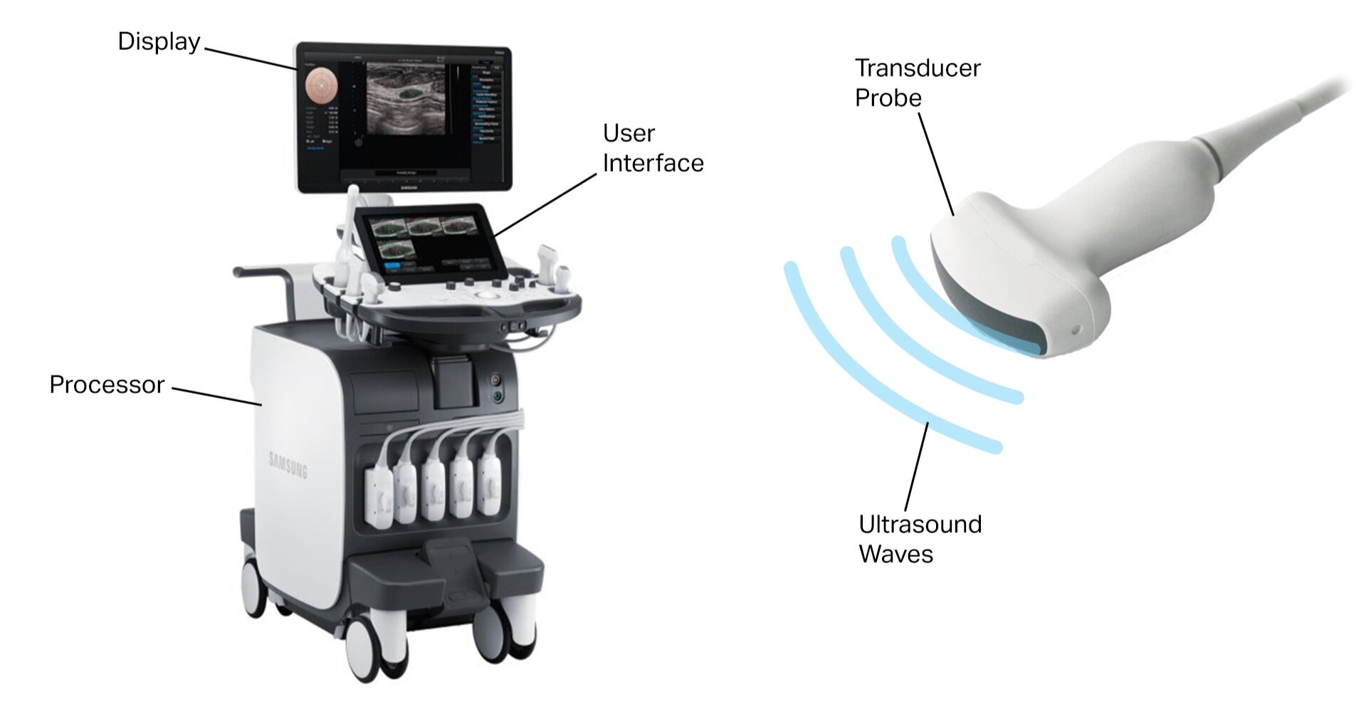
Components of a Typical Ultrasound System
Who
Some of the main companies who make ultrasound systems include:
Samsung (Crystal Architecture, CrystalBeam, CrystalPure, HERA, Prestige, EVO, Elite, Prime, Plus)
Fujifilm (Sonosite)
GE (LOGIQ, Vscan, Venue, Vivid, Versana, Voluson, ABUS)
Philips (EPIQ, Affiniti, Sparq, Xperius)
Siemens (ACUSON)
Canon, Toshiba (Aplio, Xario)
Hitachi (Precision, LISENDO, ARIETTA, ProSound)
References
http://www3.gehealthcare.com/en/products/categories/ultrasound
http://www.samsung.com/global/business/healthcare/healthcare/ultrasound
http://www.physics.utoronto.ca/~jharlow/teaching/phy138_0708/lec04/ultrasoundx.htm